2023 Scientific Meeting Poster Presentations with Abstracts

Reviewed & Presented by the AAOP Poster Committee


Poster 1

Insomnia is Associated with Higher Pain Intensity in Orofacial Pain Patients: A Cross- Sectional Study

Authors: Alessandri-Bonetti, A1,2; Sangalli, L3; Boggero, IA1,4

Affiliations: 1University of Kentucky, College of Dentistry, Department of Oral Health Science, Division of Orofacial Pain; Lexington, KY, USA. 2 Institute of Dental Clinic, A. Gemelli University Policlinic IRCCS, Catholic University of Sacred Heart; Rome – Italy. 3 College of Dental Medicine – Illinois, Midwestern University; Downers Grove, IL, USA. 4 University of Kentucky, Department of Psychology; Lexington, KY, USA

Aim of investigation: Good sleep quality is key factor underlying pain control. Yet, few studies have investigated the relationship between pain intensity and insomnia in orofacial pain patients. Our hypothesis was that insomnia symptomatology would be associated with pain intensity. Aims of the study were to assess associations of insomnia symptomatology with pain interference, pain duration, and general medical health in a population of treatment-seeking orofacial pain patients.

Methods: A cross-sectional study was conducted in consecutive adults seen at Orofacial Pain Clinic (University of Kentucky). Demographics, insomnia symptoms (assessed via Insomnia Severity Index, ISI), pain duration, pain intensity and pain interference (Graded Chronic Pain Scale) were extracted. T-tests were used to assess differences in outcomes between patients with and without insomnia.

Results: Of 272 patients (43.1±15.3 y/o, 83% females), 47.8% present with clinically significant insomnia symptomatology, defined by scores of ISI ≥ 11. Compared to patients with elevated insomnia symptoms, those without clinically elevated insomnia symptomatology (52.2%) reported lower pain intensity (44.7±20.8 vs 60.4±20.6, t=-6.158, p<.001), lower pain interference (16.5±20.2 vs 39.9±29.4, t=-7.475, p<.001) and fewer medical diagnosis (4.4±5.3 vs 6.1±5.6, t=-2.505, p<.01). No significant difference was observed in mean pain duration (75.6 vs 79.7 months, p>.05) between the two groups. Group differences remained significant after adjusting for age, sex, and primary orofacial pain diagnosis.

Conclusion: Patients with insomnia experienced higher pain intensity, greater pain interference and poorer general health than patients without insomnia, thus highlighting the importance of sleep examination in orofacial pain patients.

Acknowledgment and Funding Source: None.

Poster 2

Nurses and Teachers are More Frequently Seen among Patients with Chronic Orofacial Pain. A Retrospective Study

Authors: Sangalli, L1; Alessandri-Bonetti, A2; Boggero, IA2,3

Affiliation: 1 College of Dental Medicine – Illinois, Midwestern University, Downers Grove, IL, USA. 2 Department of Oral Health Science, Division of Orofacial Pain, University of Kentucky, College of Dentistry, Lexington, KY, USA. 3 Department of Psychology, University of Kentucky, College of Arts and Science, Lexington, KY, USA

Aim of investigation: Occupational status may be related to how people experience and seek care for orofacial pain (OFP). Yet, no study has investigated the prevalence of specific job categories among treatment-seeking patients with OFP at a tertiary clinic, which constituted the aim of this study. A secondary aim was to describe employment status and test associations with clinical, sleep, or psychological variables.

Methods: A chart review was conducted for consecutive adults seen at OFP Clinic (University of Kentucky). Demographics (age, sex, employment status, profession), insomnia symptoms (ISI), anxiety-depression (PHQ-4) and pain intensity (GCPS) were extracted. One-way ANOVA was used to test differences in outcomes among different working status.

Results: Of 333 patients (43.8±15.9 y/o, 82% females), 7.6% were students, 64.5% employed, 11.6% retired, 7.0% disabled, 9.3% unemployed. The two most common jobs were nurses (5.6%) and teachers (7.0%). All the other job categories had frequencies <1.5%. Teachers exhibited less anxiety-depression compared to the other jobs (2.6±2.5 vs 4.06±3.1, p=.050). No difference was observed in any other variables among job categories. Those who were working had significantly worse insomnia symptomatology (11.7±7.2 vs 9.8±6.2, p=.018), greater anxiety-depression (4.7±3.4 vs 3.8±3.1, p=.031) and poorer sleep quality (8.8±4.3 vs 7.6±3.9, p=012) than those who were working. Unemployed patients exhibited greater pain intensity than employed (p=.047) and disabled patients (p=.001).

Conclusion: Nurses and teachers were overrepresented among employed patients seeking treatment for chronic orofacial pain. Future studies should explore the job characteristics (i.e., high demand, environment, extensive talking) that predispose certain professions to orofacial pain.

Acknowledgment and Funding Source: None.

Poster 3

Are Educational Blogs a Suitable Strategy to Promote Awareness About Orofacial Pain- Related Topics? A 4-Year Outcome Report

Authors: Wang, JC; Botros, J; Padilla, M

Affiliations: Herman Ostrow School of Dentistry, University of Southern California, Los Angeles, CA

Aim of investigation: To determine if open-source educational blogs can help promote orofacial pain specialty awareness.

Methods: Viewership data of the USC Ostrow online webpage analytics for the top-viewed orofacial pain and pain medicine blogs published between 12/2018 and 12/2022 was collected. The percentage of returning visitors, the number of days since publication and the number of words per blog were calculated. Blogs were categorised by content (examination/diagnostic, treatment and comprehensive), and the impact of the pandemic was factored using a chronological analysis of the data. Multivariate linear regression models adjusted for relevant covariates were employed.

Results: A total of 85 blogs were analyzed, with a mean view count of 577 pre-pandemic, 1,490 during the 1st wave, and 3,338 after the 1st wave. The mean time spent per blog was longer during (p=0.04) and after (p<.001) the 1st wave compared to pre-pandemic. The percentage of blog re- visitors decreased both during and after the 1st wave. Since the blogs stay in the webpage, the number of views correlates with the posted time. Treatment-focused blogs were more frequently viewed than comprehensive ones (p=0.04).

Conclusions: This study indicated that the use of a content-based strategy with open-source blogs has the potential to generate interest in orofacial pain-related topics, which increases with posted time. Open-source educational blogs should be considered as a strategy to expand awareness of the new dental specialty of orofacial pain. Research should be conducted to determine if blog readership spiked in other similar disciplines.

Acknowledgements and Funding Source: None.

Poster 4

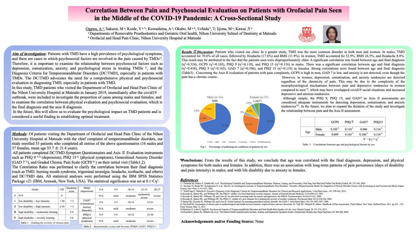
Correlation Between Pain and Psychosocial Evaluation on Patients with Orofacial Pain Seen in the Middle of the COVID-19 Pandemic: A Cross-Sectional Study

Authors: Ogawa, A; Sakurai, M; Koide, Y; Kuwashima, A; Okubo, M; Uchida, T; Iijima, M; Kawai,Y

Affiliations: Departments of Removable Prosthodontics and Geriatric Oral health, Nihon University School of Dentistry at Matsudo

Aim of investigation: The purpose of this study was to assess pain and psychosocial evaluation using DC/TMD instruments in patients with orofacial pain seen in the middle of the COVID-19 pandemic.

Methods: Fifty-three patients (16 males and 37 females, mean age 53.7 ± 21.4 years) who presented to the Orofacial and Head Pain Clinic at NUSDM Hospital with a chief complaint of orofacial pain participated. All patients completed DC/TMD symptom questionnaires and Axis II evaluation instruments such as PHQ9 (depression), PHQ15 (physical symptoms), GAD7 (Generalized Anxiety Disorder), and GCPS (Graded Chronic Pain Scale) on their initial visit. Multivariate analysis (One-way ANOVA, Correlation Ratio, SPSS) was performed to clarify the correlation between their final diagnosis (such as temporomandibular disorders, burning mouth syndrome, trigeminal neuralgia, headache, toothache, and others) and DC/TMD data.

Results: A significant correlation was found between age and final diagnosis (eta2=0.324), GCPS (eta2=0.105), PHQ9 (eta2=0.118), and PHQ15 (eta2=0.116) in males. There was a significant correlation between age and final diagnosis (eta2=0.438), PHQ9 (eta2=0.165), GAD7 (eta2=0.106), and PHQ15 (eta2=0.110) in females. Particularly strong correlations were found between age and final diagnosis.

Conclusions: From the results of this study, we conclude that age was correlated with final diagnosis, depression, and physical symptoms for both men and women. In addition, there was an association with long-term patterns of pain persistence (days of disability and pain intensity) in men, and with life disability due to anxiety in women.

Acknowledgements and Funding Source: None.

Poster 5

Management of Concomitant Trigeminal Neuropathy and Myofascial Pain with Botulinum Toxin-A (BoNT-A)

Authors: Kandari, A; Sawani, S; Hernandez, I; Friesen, R

Affiliations: TMD/Orofacial Pain Clinic (Oral Medicine Graduate Program) University of Alberta, Canada

Aim of investigation To stress that correct diagnoses are critical for successful pain management.

Methods: A 26-year-old female complained of persistent pain in quadrant 1. The onset of this pain followed a root canal treatment on tooth #4. Prior to that, she experienced discomfort in teeth # 2, 3, 4 and 5. She had been assessed by the family dentist, a prosthodontist, and an endodontist without reaching a definitive diagnosis. She had been fitted with a maxillary splint and advised physiotherapy. Upon our assessment, the patient reported continuous moderate to severe recurring, shooting paroxysms of pain with burning sensation. Mastication and cold air triggered the pain. There were trigger points associated with the right masticatory musculature. Percussion of the affected teeth exacerbated the pain for more than 12 hours.Our diagnoses were trigeminal neuropathy and myofascial pain. Previously acquired brain MRI ruled out central abnormalities. We discussed our findings with the patient, prescribed baclofen for its benefits on neuropathic and muscular pain, recommended continuation of physiotherapy and fabricated a mandibular splint.

Results: The patient reported a lesser severity only in the muscular pain. We proceeded with nerve blocks in the affected teeth and trigger point injections with complete but short-lived pain relief. We then used botulinum toxin-A (BoNT-A) for the neuropathic and myofascial pains. Botulinum toxin injections resulted in near-complete resolution of pain. This ongoing pain management has been effective for three years now.

Conclusions: Diagnosing and managing the concomitant neuropathic and myofascial pains was essential for optimal pain management.

Acknowledgements and Funding Source: None.

Poster 6

The DDS and ASPID Students’ Perspective Regarding the “Virtual Flipped Classroom, Case- Based” Pedagogy Introduced Through Orofacial Pain Module: A Quantitative Survey Study

Authors: Ahmadieh, A1 and Navazesh, M2

Affiliations: 1Clinical Assistant Professor, Herman Ostrow School of Dentistry of USC and Diplomate of American Board of Orofacial Pain. 2Professor and Associate Dean, Herman Ostrow School of Dentistry of USC

Aim of investigation To stress that correct diagnoses are critical for successful pain management.

Methods: A 26-year-old female complained of persistent pain in quadrant 1. The onset of this pain followed a root canal treatment on tooth #4. Prior to that, she experienced discomfort in teeth # 2, 3, 4 and 5. She had been assessed by the family dentist, a prosthodontist, and an endodontist without reaching a definitive diagnosis. She had been fitted with a maxillary splint and advised physiotherapy. Upon our assessment, the patient reported continuous moderate to severe recurring, shooting paroxysms of pain with burning sensation. Mastication and cold air triggered the pain. There were trigger points associated with the right masticatory musculature. Percussion of the affected teeth exacerbated the pain for more than 12 hours.Our diagnoses were trigeminal neuropathy and myofascial pain. Previously acquired brain MRI ruled out central abnormalities. We discussed our findings with the patient, prescribed baclofen for its benefits on neuropathic and muscular pain, recommended continuation of physiotherapy and fabricated a mandibular splint.

Results: The patient reported a lesser severity only in the muscular pain. We proceeded with nerve blocks in the affected teeth and trigger point injections with complete but short-lived pain relief. We then used botulinum toxin-A (BoNT-A) for the neuropathic and myofascial pains. Botulinum toxin injections resulted in near-complete resolution of pain. This ongoing pain management has been effective for three years now.

Conclusions: Diagnosing and managing the concomitant neuropathic and myofascial pains was essential for optimal pain management.

Acknowledgements and Funding Source: None.

Poster 7

Agenesis of the Temporomandibular Condyle in a Three-Year-Old Patient: A Case Report

Authors: Benavides, S1 and Pérez, C2

Affiliations: 1Resident Orofacial Pain. University of Kentucky, Lexington. 2Associate Professor and Chief, Division of Pediatric Dentistry, University of Kentucky, Lexington

Aim of investigation: Complete absence of the temporomandibular condyle is an extremely uncommon condition, especially in a healthy child. This condition may not be noticed at an early age, however as the child grows it can manifest with pain, facial asymmetry, and functional disturbances. This work presents a rare case of left condylar aplasia/agenesis in a young child.

Methods: A 3-year-old girl presented to the Orofacial Pain Center at the University of Kentucky with pain and dysfunction located in the left masseter muscle, pre-auricular area and ear. A thorough anamnesis, clinical examination and imaging were performed. A panoramic radiograph and subsequent CT scan showed a lack of development of the left mandibular ramus and complete absence of the left temporomandibular condyle.

Results: The patient was referred to plastic surgery where the diagnosis of left condylar agenesis/aplasia was confirmed, and distraction osteogenesis was indicated as future treatment. Other management options were discussed by orthodontics and pediatric dentistry including functional appliances that may guide growth and avoid invasive surgery.

Conclusions: The presence of a deviated chin, tilting of the mandibular plane and facial pain in a healthy young child can be early manifestations of a skeletal anomaly relating to the temporomandibular joint. Prompt recognition is essential as it provides reassurance to the parents and initiates the consideration of treatment options. Making an early diagnosis as well as initiating early treatment that employs the child’s growth potential may avoid further facial asymmetry and pain.

Acknowledgements and Funding Source: None.

Poster 8

Myositis Ossificans of the Masseter Muscle – A Case Report

Authors: Alswaiti, O1 and Malacarne, A2

Affiliations: 1 Orofacial Pain Resident, Department of Diagnostic Sciences, School of Dental Medicine, Tufts University, USA. 2 Assistant Professor, Department of Diagnostic Sciences, School of Dental Medicine, Tufts University, USA

Aim of investigation: In this case report we describe a rare case of myositis ossificans of the masseter muscle. A 33-year-old female, already treated for chronic orofacial pain at our clinic diagnosed as fibromyalgia, presented with an acute complaint of left side jaw swelling and limited mouth opening. Patient reported falling and hitting the left side of her face on a sidewalk two months prior to our visit. She developed acute swelling, bruising and increased jaw pain. Swelling reduced, bruising resolved, and patient currently reports facial asymmetry, a lump on the left cheek and inability to fully open her mouth.

Methods: Head and neck examination revealed a hard lump in the body of the masseter muscle, slightly tender to touch. Maximum mouth opening is 25 mm with hard end-feel. A CT scan reveals no fracture and the presence of a 1.0 x 1.7 mm well-defined, irregular, calcified lesion in the body of the left superficial masseter.

Results: Patient was diagnosed with myositis ossificans of the left masseter, she was referred to oral and maxillofacial surgery for a consultation. The joint treatment plan consists of observation for 3 months for potential spontaneous remission, followed by surgical excision and biopsy of the lesion. Post-surgical physical therapy is recommended to prevent masseter fibrotic contracture.

Conclusion: Chronic orofacial pain presenting with new complaints should be thoroughly assessed to differentiate between exacerbation of her chronic symptoms versus new onset diagnoses. Myositis ossificans, despite rare, should be included in the differential diagnosis in the presence of trauma and hard swelling.

Acknowledgements and Funding Source: None.

Poster 9

Clinical Reasoning Process in Patients with Temporomandibular-Related Somatic Tinnitus: A Case Series

Authors: van der Wal, AC 1,2,3; Visscher, CM 1; van der Meer, HA 1,3; De Hertogh, W2

Affiliations: 1 Department of Orofacial Pain and Disfunction, Academic Centre for Dentistry Amsterdam (ACTA), University of Amsterdam and Vrije Universiteit (VU) University Amsterdam, Amsterdam, the Netherlands. 2 Department of Rehabilitation Sciences and Physiotherapy, Faculty of Medicine and Health Sciences, University of Antwerp, Antwerp, Belgium. 3 SOMT University of Physiotherapy, Amersfoort, the Netherlands

Aim of investigation: Patients with tinnitus resulting from temporomandibular dysfunction increasingly present themselves in orofacial clinics. Despite growing evidence showing positive effects for TMD-treatment on tinnitus, these patients remain a challenge to manage.
Five cases with somatic tinnitus with a detailed description about the clinical reasoning process and applied treatment are presented.

Methods: A case series presenting three women and two men (mean age:49). An extensive tinnitus and TMD assessment were performed in an orofacial physical therapy clinic. The assessment consisted of medical history (age, gender, duration of tinnitus), otologic symptoms (hearing loss, type of tinnitus) and TMD (according to the DC-TMD). Additionally, physical examination of the neck was completed. Treatment was personalized according to findings from the clinical examination and consisted of counseling, habit reversal techniques, relaxation exercises, mobilization techniques and massage/stretching of the jaw and neck muscles.

Results: According to the diagnostic criteria of somatic tinnitus, the patients had: modulation of tinnitus by jaw movement (n=1), modulation by pressure on sternocleidomastoid (n=2), jaw complaints and tinnitus appeared simultaneously (n=1), tinnitus pitch and location vary (n=3), TMD myalgia (n=4), bruxism (n=5), neck pain (n=4) and hearing loss (n=1). Tinnitus duration was on average three years. The orofacial physical therapy took between eight and eleven sessions applied within three months. All patients noticed a reduction of their tinnitus as measured by the global perceived effect score.

Conclusions: The cases presented provide better insight in the clinical reasoning process and treatment in patients with somatic tinnitus.

Acknowledgements and Funding Source: None.

Poster 10

Application of Bayesian Inference Algorithm in Orofacial Pain and Oral Medicine Diagnoses: A Retrospective Study

Authors: Vistoso Monreal, AP1; Veas, NA2; Loeb GE3; Clark, GT1

Affiliations: 1Herman Ostrow School of Dentistry, University of Southern California. 2McCombs School of Business, The University of Texas. 3Viterbi School of Engineering, University of Southern California

Aim of investigation: Bayesian inference is an Artificial Intelligence algorithm based on Bayes’ theorem that successfully uses probabilities to make predictions. We aim to test the integration of this algorithm into the Smart Note (SN), a customized highly structured note-taking system of the Orofacial Pain (OFP) and Oral Medicine (OM) Center of USC. The implementation of this diagnostic support system is to improve decision-making and accuracy in OFP and OM diagnoses.

Methods: Bayesian Algorithm was trained with retrospective data of 1020 clinical notes of patients seen between July 2021-Nov 2022 at the OFP and OM center of USC. Phyton library was used for statistical analysis and implementation of the algorithm.

Results: 20 random cases were selected to evaluate the performance of the algorithm. Of 115 diagnoses (OFP+OM) registered in the system, the algorithm could predict the diagnoses with 98.5% accuracy and 59% precision. The ability to predict a true positive diagnosis was 61% and a true negative was 60%.

Conclusions: We demonstrated the feasibility of the implementation of the Bayesian inference algorithm into a note-taking system to support decision-making in OFP-OM disciplines. Future analysis with real-time patients and a much large dataset will validate the acceptance and performance of the algorithm implemented in the SN.

Acknowledgments and Funding Source: None.

Poster 11

Inhibition of Nociception in a Preclinical Chronic TMD Model by Dietary Supplementation with Grape Seed Extract is Associated with Increased Expression of GABAergic Proteins in the Trigeminal Ganglion

Authors: Garten, DA; Antonopoulos, SR; Woodman, SE; Durham, PL

Affiliation: Missouri State University, Springfield, MO

Aim of investigation: To investigate cellular changes in protein expression in the trigeminal ganglion and spinal trigeminal nucleus in a chronic TMD model and in response to daily supplementation with grape seed extract.

Methods: Mechanical nociception was determined using von Frey filaments and cellular changes in protein expression evaluated by immunohistochemistry in trigeminal ganglion and spinal trigeminal nucleus. The three experimental conditions included naïve, animals subjected to neck muscle inflammation and prolonged jaw opening, and daily supplementation with grape seed extract in the drinking water prior to inducing TMD pathology.

Results: The mechanical sensitivity observed in the TMD model was significantly suppressed by inclusion of the extract. In the trigeminal ganglion, neuronal levels of GAD65/67 and GABAB, which are proteins involved in inhibiting sensitization and activation of trigeminal neurons, were decreased in the TMD model when compared to control animals. Importantly, supplementation with the extract promoted a significant increase in the expression of those proteins. In the trigeminal spinal trigeminal nucleus, increased expression of GFAP, a biomarker of activated astrocytes, was observed in the TMD model. However, the elevated levels of GFAP were not suppressed by dietary inclusion of the extract.

Conclusion: Our findings provide evidence of a neuroprotective role of grape seed extract in the trigeminal ganglion and support the notion that the inhibitory effect of the extract involves, in part, increased neuronal synthesis of the inhibitory neurotransmitter GABA and expression of GABAB receptor in neurons and glia to suppress peripheral sensitization.

Acknowledgements and Funding Source: Missouri State University.

Poster 13

Apnea-Hypopnea Index (AHI) and Oxygen Desaturation in Obstructive Sleep Apnea (OSA) Patients: A Retrospective Study

Authors: Masoudian Khouzani, M; Venturin, J; Sheppard, G; Sedghizadeh, P; Clark, G; Padilla, M

Affiliation: Orofacial Pain and Oral Medicine Clinic, University of Southern California

Aim of investigation: To examined oxygen desaturation characteristics of patients referred to the Orofacial Pain and Oral Medicine (OFPM) Clinic with a diagnosis of obstructive sleep apnea (OSA).

Methods: Retrospective study conducted at the University of Southern California (IRB# UP-07- 00416), with data collected from 9/2015 to 7/2021. A single researcher reviewed all the charts of patients diagnosed with OSA and randomly selected 49 (N=101). All data was analyzed using descriptive statistics.

Results: The data suggests a positive correlation between high blood pressure and AHI (Apnea- Hypopnea Index), which is not statistically significant (correlation = 0.095, p = 0.527). A significant negative correlation was found between AHI and SPO2 nadir (correlation = -0.257, p- value of 0.075). This suggests that as OSA severity increases, minimum oxygen saturation levels decrease. A negative correlation was found between AHI and SPO2 average, but this was not statistically significant (correlation = -0.233, p = 0.107). The data showed no significant differences in average AHI when comparing genders (p = 0.631). This sample also shows that as age increases so does AHI (rho=0.174; p = 0.233) though this was not statistically significant, however, SPO2 average decreases significantly with age (rho=-0.287; p = 0.045), as well as SPO2 nadir (rho=- 0.488; p <= 0.001).

Conclusion: Patients with OSA should be monitored for high blood pressure, and older adults should be assessed for sleep disorders. Further studies with bigger samples are needed to confirm these findings.

Acknowledgements and Funding Source: Dr. Reyes Enciso contributed with the statistical analysis.

Poster 14

Squamous Cell Carcinoma Misdiagnosed and Treated as a Masticatory Muscle Disorder: A Case Report

Authors: Mishra, SS1; Guerrero, P2; Correa, L3

Affiliations: 1Resident, Department of Diagnostic Sciences, Tufts University School of Dental Medicine. 2Assistant Professor, Department of Diagnostic Sciences, Tufts University School of Dental Medicine.3Associate Professor, Department of Diagnostic Sciences, Tufts University School of Dental Medicine

Aim of investigation: In this case report we describe a case of oral squamous cell carcinoma that was initially misdiagnosed as a masticatory muscle disorder by a dentist and was later treated by a TMD specialist. A 78-year-old male, presented with limited mouth opening accompanied by dull, yet sometimes sharp pain in the pre-auricular region. He reported losing 12-14 lbs in the 6 months related to “trouble chewing”. He allegedly saw two dentists who provided him with botulinum toxin injections, oral appliance, and muscle relaxants, all of which were unsuccessful in improving symptoms. Medical history revealed smoking and multiple basal cell carcinomas of his face.

Methods: Examination revealed 10 mm of maximum mouth opening with a palpable firm mass noted intraorally along the right ascending ramus in the same region the panoramic x-ray which exhibited significant destruction of bone. Mild pain was noted upon palpation of right masseter and sternocleidomastoid muscles. Bilateral lymphadenopathy was detected in upper cervical region. A referral to the Department of Oral and Maxillofacial Surgery was made to perform a biopsy of the lesion in question.

Results: Final diagnosis was given as basaloid squamous cell carcinoma. The patient is being followed up by a multidisciplinary team of oral surgeons, oncologist, and ENT specialists at Tufts Medical Center.

Conclusions: Accurate diagnosis requires careful history taking, complete examination, and multidisciplinary management. Precise identification of the source of pain is a key to correct diagnosis, prevention of diagnostic delay and unwarranted treatment options.

Acknowledgements and Funding Source: None.

Poster 15
Relationship between Migraine and Myofascial Orofacial Pain: A Systematic Review

Authors: Iturriaga, V1; Velásquez, N2; Bornhardt, C1; Thomas, DC4

Affiliations: 1Assistant Professor, Department of Integral Adult Care Dentistry, Temporomandibular Disorder and Orofacial Pain Program, Sleep & Pain Research Group, Faculty of Dentistry, Universidad de La Frontera, Temuco, Chile. 2Temporomandibular Disorder and Orofacial Pain Program, Faculty of Dentistry, Universidad de La Frontera, Temuco, Chile. 3Assistant Clinical Professor, Rutgers School of Dental Medicine, Newark, USA

Aim of investigation: The aim of this systematic review was to assess the relationship between migraine and myofascial orofacial pain (MOFP).

Methods: A systematic review was conducted in MEDLINE, WoS, SCOPUS databases, and search engine Trip Database. References of the included articles were reviewed to identify other publications. The inclusion criteria were observational studies and controlled clinical trials that assessed the relationship between migraine and MOFP in humans, limiting the search to studies published in the last 10 years, either in English or Spanish.

Results: The initial search yielded 345 articles, 10 were finally included in the analysis. Migraine patients have a prevalence of MOFP between 38% and 44%. Both pathologies share the same nociceptive system, headed by the caudal nucleus of the trigeminal nerve, the trigeminal-cervical complex and the trigeminal-vascular system. Also, both entities are related to central and peripheral sensitization of the trigeminal nerve, generating abnormal nociceptive processing. Comorbidity between them is bidirectional, migraine could be a risk factor for the development of MOFP, and MOFP may favor the onset or subsequent chronification of episodic migraine to chronic migraine.

Conclusions: Results of this systematic review suggest that there is a close relationship between migraine and MOFP, sharing neuroanatomical structures and pathophysiological mechanisms. It is essential to consider this close relationship, where the timely identification of this comorbidity could help to optimize the diagnosis and management of these patients.

Acknowledgements and Funding Source: Temporomandibular Disorders and Orofacial Pain Program and Project DI20-0018, Universidad de La Frontera.

Poster 16

Paradoxical Masseteric Bulging after Botulinum Neurotoxin Type A Injection: A Case Report

Authors: Sandhu, S and Shaefer, J

Affiliation: Mass General Hospital/Harvard School of Dental Medicine

Aim of investigation: We describe a case of paradoxical masseteric bulging after botulinum neurotoxin type A injection- a rare side effect. A 46-year-old female presented for evaluation of bilateral jaw pain. She described the pain as a constant 5-8/10 ache. She had previously tried muscle relaxers, NSAIDs, physical therapy, trigger point injections and occlusal orthotic without much benefit.

Methods: The clinical examination revealed bilateral masseteric hypertrophy with tenderness of masseters. Maximal mouth opening was 50 mm with normal lateral movements. The panoramic imaging did not reveal any degenerative condylar changes.

Results: Patient was diagnosed with chronic myofascial masticatory pain and masseteric hypertrophy in the setting of day and night-time bruxism. It was decided she would benefit from botulinum toxin injections (30 units each for masseters and 20 units each for temporalis muscles). She developed an uncommon side effect of bilateral masseteric bulging after botulinum toxin. To counter this adverse effect, 20 units each of botulinum toxin was injected into the bulged masseter areas in the subsequent visit with excellent response.

Conclusion: Paradoxical masseteric bulging, a rare sequela of botulinum toxin into hypertrophied masseter, may occur due to presence of a deep tendon structure within superficial masseter that divides it into a superficial and deep belly. This tendon confines the injected toxin to within deeper muscle belly and prevents spreading of botulinum toxin into the entire layer of the superficial muscle belly resulting in the bulging of the part of superficial belly, unaffected by botulinum toxin.

Acknowledgements and Funding Source: Education and Research Fund, MGH Oral Surgery.

Poster 17

HIV-Related Arthritis Affecting the Temporomandibular Joints: A Case Report

Authors: Taema, M1, 3; Uppgaard, R2; Herman, C1

Affiliations: 1 University of Minnesota Department of Diagnostic and Biological Sciences, Division of TMD and Orofacial Pain. 2 University of Minnesota Department of Developmental and Surgical Sciences, Division of Oral and Maxillofacial Surgery. 3 Cairo University, Department of Prosthodontics

Aim of investigation: We present a case of HIV-related arthritis affecting the temporomandibular joint (TMJ). Although approximately 5% of patients with HIV infection may develop HIV-related arthritis, HIV-related degeneration of the TMJ is an uncommon finding.
Reactive arthritis, an inflammatory process that commonly is a reaction to bacterial/viral pathology, is a known complication of HIV. To the best of our knowledge, this is the first documented case of HIV-related arthritis affecting the TMJ.

Methods: A 58-year-old male presented with a previous diagnosis of HIV, and complaints of jaw pain with limited mouth opening. MRI findings revealed an anterior disc displacement with mild inflammation. During Covid pandemic, he did not return until 2 years later with increased pain, limited mouth opening, a 5 mm anterior open bite and only molar occlusion; all of which affected his function and speech. A comparative MRI revealed inflammatory arthropathy with severe bilateral condylar degenerative changes. Serological investigations were inconclusive, however, evaluation with rheumatology revealed severe concurrent first metacarpophalangeal joint degeneration.

Results: A combined surgical and rehabilitative approach was implemented. Surgical treatment including a custom TMJ prosthesis (TMJ Concepts) was fabricated and used to restore normal occlusion and function. Post-operatively, he was admitted to physical therapy to restore adequate range of motion. To date, he has stable symptoms and has regained proper function.

Conclusion: Advanced degenerative disease of the TMJ can result in significant functional limitations and dentofacial deformity. A multidisciplinary approach with close monitoring is necessary for proper management.

Acknowledgements and Funding Source: None.

Poster 18

Atypical Vestibular Migraine and Its Response to Various Treatment Modalities: A Case Report

Authors: Mehrotra, S and Eli, B

Affiliation: Private Practitioners

Aim of investigation: This case report describes an atypical case of vestibular migraine characterized by periods of severe nausea, vomiting, and vertigo without any head or face pain that was initially diagnosed as Meniere’s disease by an ENT but did not respond to associated Meniere’s treatments. Symptoms responded favorably to onobotulinumtoxinA injections and oral appliance therapy, and failed to respond to classic migraine abortive and anti-nausea medications. It serves as an example that not all migraines fit the classic criteria nor are they always accompanied by headache symptoms.

Methods: A detailed head and neck examination and history of present illness was conducted. Various treatments were initiated over the course of 15 follow-up visits including pharmacotherapy trials, nerve blocks, onobotulinumtoxinA injections, and oral appliance therapy.

Results: The patient’s symptoms did not significantly respond to classic migraine abortive treatments but remain well controlled with quarterly onobotulinumtoxinA injections and continued oral appliance use.

Conclusions: Although this patient’s vertiginous episodes did not present as classic migraine, her response to onobotulinumtoxinA and equivocal response to classic Meniere’s treatments suggests that her symptoms were due to vestibular migraine rather than Meniere’s disease. Basic understanding of migraine characteristics and variants is important. This case report demonstrates the importance of careful history taking and frequent follow up to manage atypical presentations of facial pain.

Acknowledgements and Funding Source: None.

Poster 19

A Case Report on Post-Herpetic Neuralgia Treated with Infraorbital Nerve Blocks

Authors: Zaman, I and Handa, S

Affiliation: Massachusetts General Hospital/Harvard School of Dental Medicine

Aim of investigation: We describe a case of post-herpetic neuralgia, a neuropathic pain disorder leading to persistent facial pain. An 81-year-old female presented with a constant 8/10 deep aching and burning pain on her left cheek, nose, and upper lip – the same distribution as a previous herpes zoster virus (HZV) rash 3 months ago.

Methods: Neurosensory alteration included tingling and itchiness, with no motor deficits of the left maxillary division of trigeminal nerve (V2) dermatome. A sensory assessment of the three branches of the trigeminal nerve showed allodynia and hyperalgesia in left V2. There was no evidence of scarring or rash present from her HZV outbreak.

Results: Her diagnosis was post-herpetic neuralgia with an intolerance to gabapentin, pregabalin, and Capsaicin cream 0.075%. After trialing and failing multiple medications, she lost motivation in trying nortriptyline. We offered left infraorbital nerve blocks, which brought her pain level down from an 8/10 the first visit to a 5/10 the second visit with analgesic effects lasting 3 weeks. By her 3rd visit, she had complete resolution of pain on her left cheek. We continued nerve blocks every 2 months as they were effective in managing her pain and respected her decision of not adding any new medications to her management.

Conclusions: As orofacial pain specialists gain more recognition, an awareness of the clinical presentation as well as treatment modalities involved in facial post-herpetic neuralgia is beneficial for efficacious treatment for this vulnerable population.

Acknowledgements and Funding Source: None.

Poster 20

Parotid Gland Pain Presenting as Initial Symptom of Diabetes Mellitus: A Case Report

Authors: Koide, Y; Kuwashima, A; Ogawa, A; Sakurai, M; Okubo, M; Iijima, M; Uchida, T; Kawai, Y

Affiliation: Orofacial and Head Pain Clinic and Department of Removable Prosthodontics, Nihon University School of Dentistry at Matsudo

Aim of investigation: This case report describes a paroxysmal parotid gland pain presenting as initial symptom of diabetes mellitus.

Methods: A 41-year-old male was referred to NUSDM Hospital with a complaint of excruciating and stabbing pain in the right mandibular quadrant after the first bite, which subsided over the next several bites. It was also triggered by orange juice and soup etc., but not by water. He consulted his family dentist, who performed the endodontic treatment on tooth #31 for the diagnosis of periapical periodontitis. He did not get any improvement from the treatment. He was suspected of salivary calculus and consulted the ENT, and no abnormalities were found. He denied any history of injury or surgery.

Results: A detailed medical interview and history showed that he had not had a medical checkup for about two years. An MRI investigation of the parapharyngeal space revealed no abnormalities. A trial of carbamazepine 400 mg daily did not reduce the pain. A blood test showed a HbA1c of 13.5%. He was sent to the internal medicine clinic, and the management for diabetes was started immediately. The prescription of pregabalin 75 mg daily reduced the pain along with the diabetes management. The initial complaint completely disappeared when the HbA1c returned to normal.

Conclusions: This case report described a paroxysmal salivary gland pain attributed to diabetes mellitus. It was concluded that not only idiopathic first bite syndrome but also diabetes mellitus should be considered as differential diagnosis of unclassifiable paroxysmal parotid gland pain.

Acknowledgements and/or Funding Source: None.

Poster 21

Metro vs Rural Orofacial Pain Patients: How Are They the Same, How Are They Different? A Retrospective Study

Authors: Dowling, MC1; Yanez-Regonesi, F1; Boggero, IA1,2,3

Affiliation: 1Department of Oral Health Science, Division of Orofacial Pain, University of Kentucky College of Dentistry, Lexington, Kentucky, USA. 2Department of Psychology, University of Kentucky College of Arts and Science, Lexington, Kentucky, USA. 3Department of Anesthesia, University of Kentucky College of Medicine, Lexington, Kentucky, USA

Aim of investigation: Previous research has found that chronic pain prevalence, intensity, and disability are all influenced by urbanization status, with residents of rural communities showing worse pain-related outcomes than those from metropolitan communities. However, few studies to date have examined the associations between urbanization status and orofacial pain, specifically. The objective of the present study was to describe the metro and non-metro status among treatment- seeking patients in a tertiary orofacial pain clinic and to evaluate any differences in age, sex, and clinical pain and psychological characteristics among them.

Methods: A retrospective study was conducted on consecutive patients seen in a tertiary orofacial pain clinic from May 2010 to March 2020. According to a patient’s zip code, a rural-urban continuum code was assigned and then grouped into metro and non-metro subcategories. Data were analyzed to observe possible differences in age, sex, pain intensity and disability assessed via the Graded Chronic Pain Scale (GCPS), and anxiety and depression assessed via the four-item patient health questionnaire (PHQ-4).

Results: Results revealed that of 1,106 new treatment-seeking patients in a tertiary orofacial pain clinic, 68.0% were from metro and 31.1% from non-metro communities. Statistically significant differences observed were age, with patients from non-metro communities older (p = 0.027), and pain intensity, with a higher pain intensity in non-metro communities (p = 0.016).

Conclusions: Our data revealed statistically significant differences in age and pain intensity score with the GCPS between patients living in metro and non-metro communities seeking treatment at a tertiary orofacial pain clinic.

Acknowledgement and Funding Source: None.

Poster 22

Neuromuscular Electrical Stimulation with Dry Needling Improved Muscle Function in Chronic Bell’s Palsy: A Case Report

Author: Blanchard, E and MacPherson, KL

Affiliation: Private Practitioners

Aim of investigation: This case report describes the outcomes of a 44-year-old, cis-female patient with chronic Bell’s Palsy (>18 months) who received physical therapy with intramuscular neuromuscular electrical stimulation (NMES) via Dry Needling (DN). The initial presentation included facial dyskinesia, flaccid dysarthria, and epiphora with smiling, eating, and speech.

Methods: Detailed evaluation revealed poor motor recruitment along the zygomatic and mandibular branches of the facial nerve resulting in dyskinesia with smiling and chewing, as well as narrowing of the eye. The Sunnybrook Facial Grading System (SFGS) served as the physical performance measure for this patient. This measure is a 0-100 scale assessing facial motor function, which has previously demonstrated excellent to near-perfect reliability. After six visits of conventional Physical Therapy, the patient received six additional visits with the inclusion of NMES via DN and manual therapy.

Results: The first six visits demonstrated minimal improvement (pre:post SFGS = 29/100:29/100), but the patient’s SFGS improved from 29/100 to 56/100 points following NMES by DN with marked improvement in items related to eye closure, increased eye opening at rest, improvements in smiling, as well as the elimination of epiphora.

Conclusion: Provision of NMES with DN correlated with improved motor recruitment of the patient’s musculature. Future clinical trials are needed to provide causative evidence for the present observations.

Acknowledgments and/or Funding Source: None.

Poster 23

Changes in Hearing Threshold and Auditory Brainstem Pathway Correlate with Trigeminal Nociception in a Preclinical Chronic TMD Model

Authors: Masten, A; Woodman, SE; Kaf, W; Durham, PL

Affiliation: Missouri State University, Springfield, MO

Aim of investigation: To utilize an established preclinical model of chronic temporomandibular joint disorder (TMD) to investigate changes in hearing sensitivity by measuring neural activity of the auditory brainstem before and post TMD.

Methods: Mechanical nociception and tone-burst auditory brainstem responses (tb-ABRs) on hearing threshold, and 80 dB response amplitude and latency at several frequencies were determined in a chronic preclinical model of TMD using Sprague Dawley rats. The three experimental conditions included naïve, animals subjected to neck muscle inflammation and prolonged jaw opening, and daily supplementation with grape seed extract (GSE) prior to inducing TMD pathology.

Results: The increase in nocifensive responses in the TMD model was attenuated by dietary supplementation with GSE. At baseline, ABR wave II was most prominent at 80 dB. As frequencies increased, latencies decreased, and amplitudes increased. ABR thresholds were normal, with decreased ABR thresholds at the higher frequencies. After induction of TMD pathology, ABR changes included earlier latency, enhanced amplitudes at all frequencies, and a 5-10 dB increase in the hearing thresholds, with the greatest increases at 12 kHz and 22 kHz. Animals receiving daily supplementation of GSE had similar hearing changes to control animals.

Conclusion: Our findings provide evidence of mild changes in hearing sensitivity and faster neural conduction as shown by earlier tone-burst ABR latency and enhanced amplitude responses. Use of this preclinical chronic TMD model will facilitate testing of novel therapeutic strategies to inhibit both the pain and auditory changes associated with TMD.

Acknowledgements and Funding Source: Missouri State University.

Poster 24

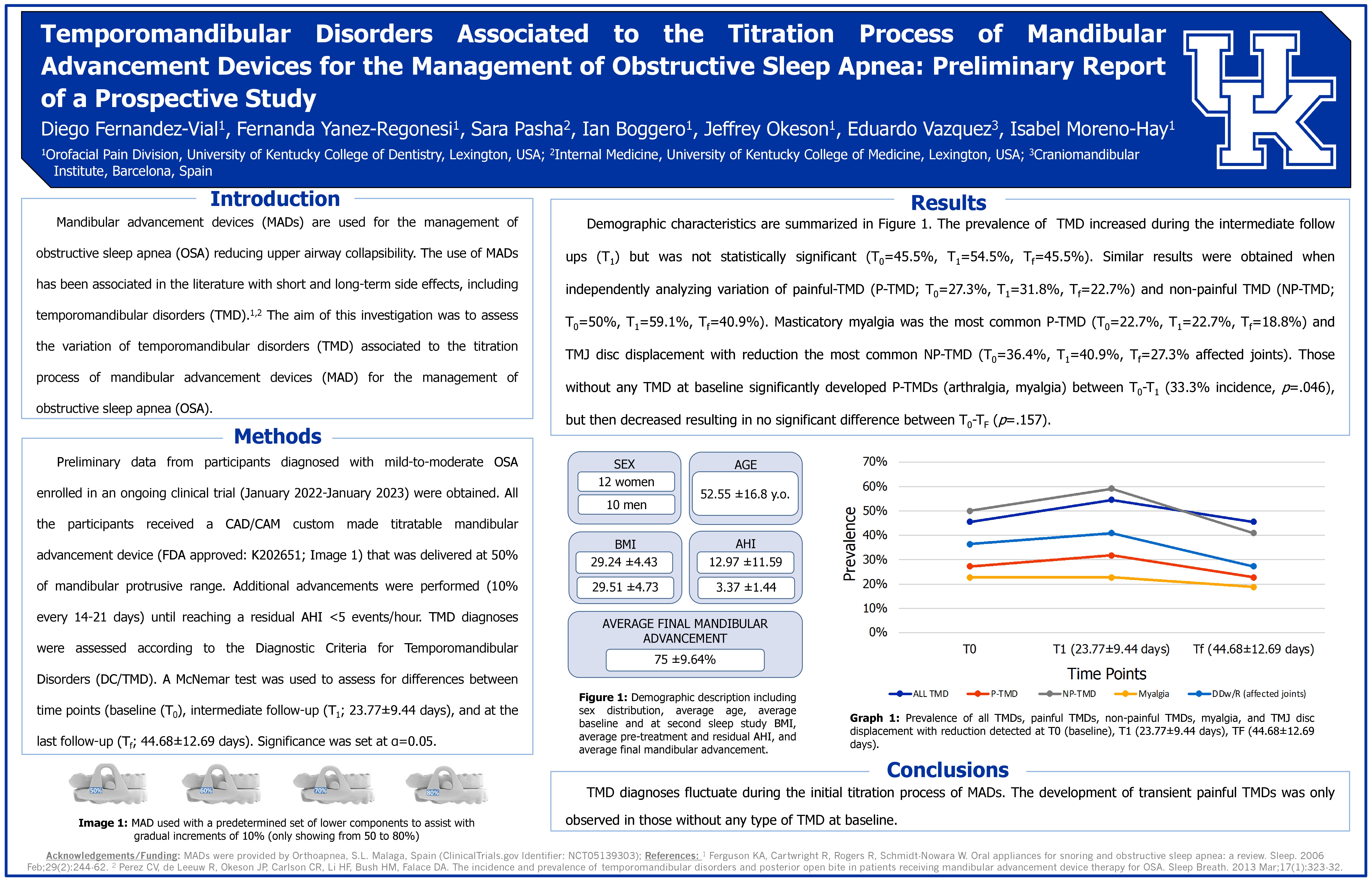
Temporomandibular Disorders Associated to the Titration Process of Mandibular Advancement Devices for the Management of Obstructive Sleep Apnea: Preliminary Report of a Prospective Study

Authors: Fernandez-Vial, D1; Yanez-Regonesi, F1; Pasha, S2; Boggero, I1; Okeson, J1; Vazquez, E3; Moreno-Hay, I1

Affiliations: 1Orofacial Pain Center, University of Kentucky College of Dentistry, Lexington, USA. 2Internal Medicine, University of Kentucky College of Medicine, Lexington, USA. 3Craniomandibular Institute, Barcelona, Spain

Aim of investigation: To assess the variation of temporomandibular disorders (TMD) associated to the titration process of mandibular advancement devices (MAD) for the management of obstructive sleep apnea (OSA).

Methods: Preliminary data from participants diagnosed with mild-to-moderate OSA enrolled in an ongoing clinical trial (January 2022-January 2023) were obtained. MAD was delivered at 50% of mandibular protrusive range. Additional advancements were performed (10% every 14-21 days) until reaching a residual AHI <5 events/hour. TMD diagnoses were assessed according to the Diagnostic Criteria for Temporomandibular Disorders (DC/TMD). A McNemar test was used to assess for differences between time points (baseline (T0), intermediate follow-up (T1; 23.77±9.44 days), and at the last follow-up (Tf; 20.91±6.63 days). Significance was set at α=0.05 for all analyses.

Results: 22 participants were included (56.5% females, 52.55±16.8 years, mean MAD advancement: 75±9.64%). The prevalence of TMD increased during the intermediate follow ups (T1) but was not statistically significant (T0=45.5%, T1=54.5%, Tf=45.5%). Similar results were obtained when independently analyzing variation of painful-TMD (P-TMD; T0=27.3%, T1=31.8%, Tf=22.7%) and non-painful TMD (NP-TMD; T0=50%, T1=59.1%, Tf=40.9%). Masticatory myalgia was the most common P-TMD (T0=22.7%, T1=22.7%, Tf=18.8%) and TMJ disc displacement with reduction the most common NP-TMD (T0=36.4%, T1=40.9%, Tf=27.3% affected joints). Those without any TMD at baseline significantly developed P- TMDs (arthralgia, myalgia) between T0-T1 (33.3% incidence, p=.046), but then decreased resulting in no significant difference between T0-Tf (p=.157).

Conclusions: OSA patients may develop transient TMD during the initial titration process of MAD.

Acknowledgements and Funding Source: MADs were provided by OrthoApnea®.

Poster 25

Perceived Origin of Pain as a Predictor of Pain Severity and Pain Descriptors: A Retrospective Study

Authors: Mitchell, A1 and Boggero, I1,2, 3

Affiliations: 1Department of Oral Health Science, Division of Orofacial Pain, University of Kentucky College of Dentistry, Lexington, USA. 2Department of Anesthesia, University of Kentucky College of Medicine, Lexington, USA. 3Department of Psychology, University of Kentucky Arts and Science, Lexington, USA

Aim of investigation: To assess whether a patient’s perceived origin of orofacial pain is associated to the perceived intensity of their pain or to number of psychological pain descriptors used to describe their pain.

Methods: As part of their initial appointment at a tertiary orofacial pain clinic, 396 patients provided data on perceived origin of pain (chewing, stress, extractions, dental procedure, motor vehicle accident, orthodontic treatment, oral surgery, blow to face/head, injection, unknown origin, and other origin), pain intensity (0-10 visual-analog scale) and psychological pain descriptors (sum of psychological pain descriptors: tiring/exhausting, sickening, fearful, or punishing/cruel, 0-4 scale). Data were extracted from their medical records for the current study. Data were analyzed using one-way ANOVA and α=0.05 was set for all analyses.

Results: Those who perceived their pain started with stress reported lower pain severity (M=5.12, SD=1.70) than those perceiving their pain started from a motor vehicle accident (M=6.75, SD=2.69, p =.043), blow to the face/head (M=6.8, SD=1.83, p=.025), or from dental procedures (M=6.66, SD=2.26, p =.010). Additionally, those who reported their pain started from a motor vehicle accident (M=1.00, SD=1.28) used more psychological descriptors than those who perceived their pain started from chewing (M=0.26, SD=0.57, p=.002), orthodontic treatment (M=0.20, SD=0.42, p=.008), unknown origin (M=0.42, SD=0.69, p=.006), stress (M=0.42, SD=0.58, p=.020, and other origin (M=0.33, SD=0.80, p=.004).

Conclusions: When chronic orofacial pain is perceived to start from a motor vehicle accident, it may be associated with greater pain intensity and psychological distress than when pain starts from another origin.

Acknowledgements and/or Funding: None.

Poster 26

Management of Nummular Headache using the CGRP Inhibitor Galcanezumab: A Case Report

Authors: Mitchell, A1 and Moreno-Hay, I1

Affiliation: 1University of Kentucky College of Dentistry, Department of Oral Health Science, Division of Orofacial Pain; Lexington, KY, USA

Aim of investigation: We detail a case of nummular headache and the introduction of galcanezumab for its management. Nummular headache is a coin-shaped headache of mild to moderate or sometimes severe intensity. It can present anywhere on the scalp, but is typically present in the parietal area. Nummular headache is often refractory to treatment and can be challenging to manage for both patients and clinicians.

Methods: A 59-year-old male presented to the University of Kentucky Orofacial Pain Center with the chief complaint of “TMJ/Headaches”. He reported struggling for over 20 years with pain and disability associated with the headaches. He detailed a long history of failed treatments and many different providers including pharmacotherapy with TCA, gabapentin, botulinum toxin injections, etc. He described his headache as being “nickel” shaped and located in the right temporalis area. The pain was reported to be 7-8/10 intensity on average with a duration of up to 12 hours each day. Extraoral examination revealed pain upon palpation in a well-circumscribed circular area of pain in the right temporal area with no observed lesions. A brain MRI with and without contrast was requested to rule out any intracranial pathology.

Results: A diagnosis of nummular headache was made and the patient was treated with galcanezumab. An initial loading dose of 240 mg was prescribed and administered. The patient reported 60% improvement in pain 16 days after the loading dose. A 120 mg dose was administered 1 month later.

Conclusion: This case illustrates the potential value of galcanezumab in the treatment of nummular headache.

Acknowledgments and Funding Source: None.

Poster 27

Treatment of Invasive Candida Osteomyelitis of the Jaw: A Case Report

Authors: Park, HJ1; Choi, HI2; Jeong, SR3; Ryu, JW1

Affiliations: 1Department of Oral Medicine, School of Dentistry, Chosun University, Gwangju 61452, Republic of Korea. 2Department of Oral and Maxillofacial Surgery, Chosun University Dental Hospital, Gwangju . 3 Chosun University 61452, Republic of Korea Department of Orthodontics, School of Dentistry, Gwangju 61452, Republic of Korea

Aim of investigation: In this case, an 81-year-old male was diagnosed with maxillary sinusitis at an ear-nose-throat (ENT) and finally diagnosed with osteomyelitis caused by invasive candida due to the destruction of the jaw at oral and maxillofacial surgery (OMFS).

Methods: On computed tomography, the sinus of the right side was radiopaque, and bone destruction up to the right maxilla and anterior nasal spine was observed. ENT performed a combined operation after consultation with the department of OMFS. On sequestrectomy, perforation was observed from the sinus to the oral cavity, and a foreign body, like a fungal ball, was discharged with the sequestrum. In a two-week follow-up, bone destruction in the lingual part of the mandible was discovered with foreign bodies like the fungus ball discharged from the posterior mandible.

Results: As a result of the microbiological culture, Candida albicans and Enterococcus faecium group D were identified. Therefore, the final diagnosis was osteomyelitis caused by invasive candida. The surgical curettage of the lesion and anti-fungal drug medication was performed. Two months later, CT confirmed that bone destruction of the mandible did not proceed any further, and the patient showed good healing clinically.

Conclusions: In conclusion, candida osteomyelitis has been considered a rare infectious disease with high mortality and fatality rates. When candida osteomyelitis of the jaw is clinically suspected, prompt diagnosis, surgical intervention, and appropriate antifungal agents should be performed.

Acknowledgment and Funding source: This study was supported by research fund from Chosun University Dental Hospital, 2023

Poster 28
Blast-Induced Orofacial Pain in a Rodent Combat and Operational Stress Model

Authors: Priess, MR1; Garza, TH1; Trevino, AV1; Szczesniak, A1; McCloskey, MP1; Greene, WA1; Clifford, JL1; VandeVord, PJ2; Stark, TR1; and Urban, MJ1

Affiliations: 1U.S. Army Institute of Surgical Research, JBSA Fort Sam Houston, TX. 2Center for Injury Biomechanics, Virginia Polytechnic Institute and State University, Blacksburg, VA

Aim of investigation: Blast can cause rapid, widespread tissue damage, inducing a complex mix of nociceptive, neuropathic, and neurovascular (headache) pain, sensory hypersensitivity and impairment, sleep disturbance, and post-concussive symptoms. Stress may also cause changes to the body that worsen traumatic and non-traumatic pain. This study examined stress and directional blast effects on orofacial pain in rats. We hypothesized that stress pretreatments would exacerbate blast-induced acute orofacial pain and related neuropathology when compared to the effects of stress or blast alone.

Methods: Stress treatments consisted of three overlapping sensory (auditory, olfactory, and visual) stimuli applied continuously for seven consecutive days to produce an impending threat perception in Sprague-Dawley rats. Stressed and unstressed animals were divided into anterior blast, lateral blast, and sham trauma exposure groups, anesthetized, and administered either a single blast wave exposure in the anterior (forward-facing) or lateral (side-facing) prone position or a comparable auditory stimulus only (sham) using an Advanced Blast Simulator. Pain measures were then conducted over the course of 96 hours and stress hormone and neuropathology assessments were performed postmortem.

Results: Stress and body orientation during blast exposure have distinct effects on orofacial pain, as evidenced through mechanical hyperalgesia, mechanical allodynia, photoallodynia, and chemical hyperalgesia measures. Stress hormone and histologic analyses are underway.

Conclusions: Stress and body orientation during blast wave exposure may significantly affect blast-induced acute orofacial pain in rats.

Acknowledgements and Funding Source: This work is supported through the U.S. Army Medical Research and Development Command’s Clinical and Rehabilitative Medicine Research Program (MR2121018E).

Poster 29

Use of Oral Devices in the Management of Oral Dysesthesia: A Case Series

Authors: Chirravur, P1,2,3; Vacharotayangul, P4, 5; Sroussi. H4,5; Klasser, GD6

Affiliations: 1Oral and Maxillofacial Diagnostic Sciences, UCONN Health, 2John Dempsey Hospital, 3Neag Comprehensive Cancer Center, Farmington. 4Division of Oral Medicine and Dentistry, Brigham, and Women’s Hospital, 5Department of Oral Medicine, Infection, and Immunity, Harvard School of Dental Medicine, Boston. 6Department of Diagnostic Sciences, Louisiana State University, School of Dentistry, New Orleans
Aim of investigation: The aim of this case series is to illustrate the significance and rationale for the use of oral devices as a mechanical/physical barrier and a safe approach in the management of oral dysesthesia without experiencing antagonistic effects from pharmacotherapeutic agents.

Methods: Two patients with spontaneous (right posterior mandibular area) and evoked following dental procedures (anterior hard palatal area) oral dysesthesia symptoms associated with hyposalivation, and taste changes were evaluated. A thorough history and complete head and neck examination with exclusion of oral lesions and systemic conditions were performed. These patients were previously prescribed topical clonazepam rinses. An in-office trial with application of orthodontic wax to the affected area was performed as a preliminary test justifying the fabrication of an oral device for possible symptomatic relief.

Results: These cases demonstrated significant improvement of oral dysesthesia symptoms and amelioration of pain in two patients after the fabrication of an oral device as a mechanical/physical barrier, with or without the use of medications who presented with a constellation of symptoms. These oral devices can be safely administered with no adverse events analogous to pharmacotherapy. Additionally, it has the potential to exert a placebo effect and efficacy in pain management.

Conclusions: This case series validate the advantage of fabricating an oral device as a mechanical/physical barrier to mitigate symptomatic pain and improve quality of life in patients diagnosed with oral dysesthesia. Additionally, the cost-effectiveness and ease of fabricating the oral device have been advantageous over numerous pharmacotherapeutic agents.

Acknowledgement and Funding Source: None.

ADVANCE REGISTRATION IS NOW CLOSED. YOU MAY REGISTER ON SITE AT THE GRAND FLORIDIAN ON MAY 3-7, 2023